00261
X-ray phase-contrast imaging using an x-ray HARP camera
PF, KEK* Department of Radiology, University of Tsukuba, Japan** NHK, Japan*** NHK-ES, Japan****
○Keiichi Hirano* Toshinomu Miyoshi* Noriyuki Igarashi* Soichi Wakatsuki* Tohoru Takeda** Jin Wu** Thet-Thet Lwin** Kenkichi Tanioka*** Norifumi Egami*** Misao Kubota*** Teruo Kawai****
X-ray phase-contrast imaging is a very useful technique to observe inner structures of various objects [1, 2]. To date, area detectors such as x-ray films, imaging plates and x-ray CCD cameras have been widely used for the technique. However, for further improvement of the performance of overall imaging systems, an x-ray area detector with higher sensitivity and better spatial resolution is required. One of the most promising candidates, which can meet this requirement, is an x-ray HARP camera [3]. The x-ray HARP camera was applied, for the first time, to the x-ray phase-contrast imaging.
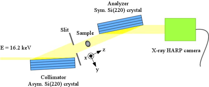
The experiment was performed at BL-14B of the Photon Factory. The experimental setup is shown below. Phase-contrast and absorption-contrast images of a rat liver were observed successfully. The sensitivity of the x-ray HARP camera was much higher than that of a fiber-coupled x-ray CCD camera. This result indicates that the x-ray HARP camera is indeed very useful for the x-ray phase-contrast imaging.
[1] A. Momose, T. Takeda, Y. Itai and K. Hirano: Nature Med., 2 (1996) 473.
[2] K. Hirano : J. Phys. D, Appl. Phys., 36 (2003) 1469.
[3] K. Tanioka and T. Hirai : OYO BUTURI (Applied Physics), 71 (2002) 1376.